基本信息
分期
複發
治療概述
恶性間皮細胞瘤治療方案
局部恶性間皮細胞瘤(I期)
進展性恶性間皮細胞瘤(II期,III期,IV期)
複發性恶性間皮細胞瘤
General Information About Malignant Mesothelioma
恶性間皮細胞瘤的基本信息
Key Points for This Section
本節要點
- Malignant mesothelioma is a disease in which malignant (cancer) cells form in the lining of the chest or abdomen.
- 恶性間皮細胞瘤是一類來源於胸部及腹部內側的恶性細胞瘤。
- Being exposed to asbestos can affect the risk of malignant mesothelioma.
- 長期暴露於石棉礦物質下將增加患恶性間皮瘤的風險。
- Possible signs of malignant mesothelioma include shortness of breath and pain under the rib cage.
- 恶性間皮瘤的癥狀包括呼吸短促及肋骨下緣疼痛。
- Tests that examine the inside of the chest and abdomen are used to detect (find) and diagnose malignant mesothelioma.
- 胸部及腹部內側檢查將有助於檢測及診斷恶性間皮細胞瘤。
- Certain factors affect prognosis (chance of recovery) and treatment options.
- 影響恶性間皮細胞瘤恢複及治療的因素
Malignant mesothelioma is a disease in which malignant (cancer) cells form in the lining of the chest or abdomen.
恶性間皮細胞瘤是一類來源於胸部及腹部內側的恶性細胞瘤
Malignant mesothelioma is a disease in which malignant (cancer) cells are found in the pleura (the thin layer of tissue that lines the chest cavity and covers the lungs) or the peritoneum (the thin layer of tissue that lines the abdomen and covers most of the organs in the abdomen). This summary is about malignant mesothelioma of the pleura.
恶性間皮細胞瘤常發生於胸膜(胸腔最內層並覆蓋肺組織)或者腹膜(腹腔最內層並覆蓋腹腔髒器),本篇主要講述發生於胸膜的恶性間皮瘤。
Respiratory anatomy; drawing shows right lung with upper, middle, and lower lobes; left lung with upper and lower lobes; and the trachea, bronchi, lymph nodes, and diaphragm. Inset shows bronchioles, alveoli, artery, and vein.
Anatomy of the respiratory system, showing the trachea and both lungs and their lobes and airways. Lymph nodes and the diaphragm are also shown. Oxygen is inhaled into the lungs and passes through the thin membranes of the alveoli and into the bloodstream (see inset).
呼吸繫統解剖圖顯示器官、肺及肺葉和氣道。還有淋巴結及隔膜。氧氣進入肺通過肺泡膜入血流。
Being exposed to asbestos can affect the risk of malignant mesothelioma.
長期暴露於石棉礦物質下將增加患恶性間皮瘤的風險
Many people with malignant mesothelioma have worked or lived in places where they inhaled or swallowed asbestos. After being exposed to asbestos, it usually takes a long time for malignant mesothelioma to form. Other risk factors for malignant mesothelioma include the following:
大部分恶性間皮瘤患者居住在或者工作在能吸入或吞入石棉換物質的地方。當暴露於石棉礦物質下時,恶性間皮瘤需要很長時間形成。恶性間皮瘤的風險因素包括:
- Living with a person who works near asbestos.
- 與接觸石棉礦物質的人生活。
- Being exposed to a certain virus.
- 長期暴露於特定的病毒。
Possible signs of malignant mesothelioma include shortness of breath and pain under the rib cage.
恶性間皮瘤的癥狀包括呼吸短促及肋骨下緣疼痛。
Sometimes the cancer causes fluid to collect around the lung or in the abdomen. These symptoms may be caused by the fluid or malignant mesothelioma. Other conditions may cause the same symptoms. Check with your doctor if you have any of the following problems:
有時腫瘤在肺和腹部產生積液,積液和恶性間皮瘤及其他狀況都將導緻以上癥狀。如果你有以下癥狀請聯繫妳的醫生:
Trouble breathing.
呼吸綑難
Pain under the rib cage.
肋緣疼痛
Pain or swelling in the abdomen.
腹痛腹脹
Lumps in the abdomen.
腹部腫塊
Weight loss for no known reason.
不明原因的體重下降
Tests that examine the inside of the chest and abdomen are used to detect (find) and diagnose malignant mesothelioma.
胸部及腹部內側檢查將有助於檢測及診斷恶性間皮細胞瘤
Sometimes it is hard to tell the difference between malignant mesothelioma and lung cancer. The following tests and procedures may be used:
有時恶性間皮瘤及肺癌很難區分。以下檢測將有助於區分:
- Physical exam and history : An exam of the body to check general signs of health, including checking for signs of disease, such as lumps or anything else that seems unusual. A history of the patient’s health habits, exposure to asbestos, past illnesses and treatments will also be taken.
- 體格及病史檢查:檢查患者的身體狀況包括疾病蹟象,如腫塊和任何異常情況。患者的生活習慣,是否暴露於石棉化合物下,病史及治療過的方案。
- Chest x-ray : An x-ray of the organs and bones inside the chest. An x-ray is a type of energy beam that can go through the body and onto film, making a picture of areas inside the body.
- 胸X攝片: 胸透X射線能穿透胸腔內器官和骨骼。x射線是一種能量束,可以穿過身體並及時使體內結構成像。
- Chest x-ray; drawing shows the patient standing with her back to the x-ray machine. X-rays are used to take pictures of organs and bones of the chest. X-rays pass through the patient onto film.
- CT scan (CAT scan): A procedure that makes a series of detailed pictures of the chest and abdomen, taken from different angles. The pictures are made by a computer linked to an x-ray machine. A dye may beinjected into a vein or swallowed to help the organs or tissues show up more clearly. This procedure is also called computed tomography, computerized tomography, or computerized axial tomography.

- CT檢查:該技術能從人體不同角度得到其內部一繫列的圖片。圖片來源於一檯與X光機器連接的電腦。病人靜脈或者口服註射造影劑,可幫助器官,或者組織更清晰的顯影。CT又被稱作計算機斷層顯像、電腦斷層攝影術或計算機化X射線軸向分層造影。
- Biopsy : The removal of cells or tissues from the pleura or peritoneum so they can be viewed under a microscope by a pathologist to check for signs of cancer.
- 活檢:從胸膜及腹膜取出腫瘤組織並在顯微鏡下觀察。
活檢方式包括以下幾種:
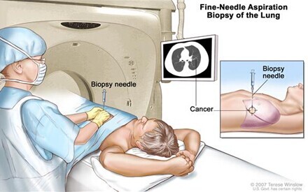
- Fine-needle (FNA) aspiration biopsy of the lung:
- 肺細針穿刺:
用細針穿刺抽取腫瘤組織或者積液,在影像引導下定位異常組織,在皮膚表面做一切口將穿刺針穿入異常組織抽取樣本。
Lung biopsy; drawing shows a patient lying on a table that slides through the computed tomography (CT) machine with an x-ray picture of a cross-section of the lung on a monitor above the patient. Drawing also shows a doctor using the x-ray picture to help place the biopsy needle through the chest wall and into the area of abnormal lung tissue. Inset shows a side view of the chest cavity and lungs with the biopsy needle inserted into the area of abnormal tissue.

Fine-Needle Aspiration Biopsy of the Lung.
The patient lies on a table that slides through the computed tomography (CT) machine, which takes x-ray pictures of the inside of the body. The x-ray pictures help the doctor see where the abnormal tissue is in the lung. A biopsy needle is inserted through the chest wall and into the area of abnormal lung tissue. A small piece of tissue is removed through the needle and checked under the microscope for signs of cancer.
肺部細針穿刺活檢:患者臥於檢查床上,CT掃描儀通過X射線成像。X射線成像圖幫助醫生發現肺部異常組織,活檢針經胸壁穿刺入肺,部分病灶抽取後置於顯微鏡下觀察腫瘤細胞。
- Thoracoscopy : An incision (cut) is made between two ribs and a thoracoscope (a thin, tube-like instrument with a light and a lens for viewing) is inserted into the chest.
- 胸腔鏡檢查:在兩肋骨間做一切借口,一細管狀帶有鏡頭和光的設備插入到胸腔內。
- Thoracotomy : An incision (cut) is made between two ribs to check inside the chest for signs of disease.
- 胸廓切開術:在兩肋骨間做一切口深入檢查胸部疾病。
- Peritoneoscopy: An incision (cut) is made in the abdominal wall and a peritoneoscope (a thin, tube-like instrument with a light and a lens for viewing) is inserted into the abdomen.
- 腹腔鏡檢查:在腹壁處做一切借口,一細管狀帶有鏡頭和光的設備插入到腹腔內。
- Laparotomy : An incision (cut) is made in the wall of the abdomen to check the inside of the abdomen for signs of disease.
- 剖腹手術:在腹壁處做一切口深入檢查腹腔內疾病。
- Open biopsy : A procedure in which an incision (cut) is made through the skin to expose and remove tissues to check for signs of disease.
- 開放性活檢:通過皮膚做一切口來探查並抽取組織進行檢查的方法。
Cytologic exam:
細胞學檢查:病理科醫生在顯微鏡下檢查異常細胞。對於恶性間皮瘤患者,將肺及腹部的積液取出並檢查其中的腫瘤細胞。
Certain factors affect prognosis (chance of recovery) and treatment options.
影響恶性間皮細胞瘤恢複及治療的因素:
The stage of the cancer.
腫瘤分期
The size of the tumor.
腫瘤大小
Whether the tumor can be removed completely by surgery.
腫瘤是否能被手術完全切除
The amount of fluid in the chest or abdomen.
胸腔及腹腔積液量
The patient's age and general health, including lung and heart health.
患者的年齡及身體狀況,包括心肺功能
The type of mesothelioma cancer cells and how they look under a microscope.
恶性間皮瘤的類型
Whether the cancer has just been diagnosed or has recurred (come back).
腫瘤是初發還是複發?
Stages of Malignant Mesothelioma
恶性間皮瘤分期
Key Points for This Section
本節要點
After malignant mesothelioma has been diagnosed, tests are done to find out if cancer cells have spread to other parts of the body.
恶性間皮瘤一旦被確診,進一步的檢查將用於判斷腫瘤是否有轉移。
There are three ways that cancer spreads in the body.
腫瘤有三種轉移方式
Cancer may spread from where it began to other parts of the body.
腫瘤可能從原發灶轉移至身體其他部位
The following stages are used for malignant mesothelioma:
恶性間皮瘤分期
· Stage I (Localized)
· I期(局部)
· Stage II (Advanced)
· II期(進展)
· Stage III (Advanced)
· III期(進展)
· Stage IV (Advanced)
· IV期(進展)
After malignant mesothelioma has been diagnosed, tests are done to find out if cancer cells have spread to other parts of the body.
恶性間皮瘤一旦被確診,進一步的檢查將用於判斷腫瘤是否有轉移。
通過觀察腫瘤是否擴散到胸膜及腹膜外可劃分分期,分期對於決定治療方案至關重要!
The following tests and procedures may be used in the staging process:
CT scan (CAT scan): A procedure that makes a series of detailed pictures of the chest and abdomen, taken from different angles. The pictures are made by a computer linked to an x-ray machine. A dye may beinjected into a vein or swallowed to help the organs or tissues show up more clearly. This procedure is also called computed tomography, computerized tomography, or computerized axial tomography.
- CT檢查:該技術能從人體不同角度得到其胸腹部一繫列的圖片。圖片來源於一檯與X光機器連接的電腦。病人靜脈註射或者口服造影劑來幫助檢測器官或者組織更清晰的顯影。該技術又被稱作計算機斷層顯像,電腦斷層攝影術或計算機化X射線軸向分層造影。
PET scan (positron emission tomography scan): A procedure to find malignant tumor cells in the body. A small amount of radioactive glucose (sugar) is injected into a vein. The PET scanner rotates around the body and makes a picture of where glucose is being used in the body. Malignant tumor cells show up brighter in the picture because they are more active and take up more glucose than normal cells do.
- 正電子掃描(正電子成像術):用於發現體內的恶性腫瘤,少量放射性葡萄糖通過靜脈註入體內,PET掃描儀旋轉於人體週圍並顯示出葡萄糖流經人體部位的圖像。圖片中恶性腫瘤細胞較正常細胞顯影明顯具有更高活性。
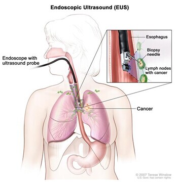
Endoscopic ultrasound (EUS): A procedure in which an endoscope is inserted into the body. An endoscope is a thin, tube-like instrument with a light and a lens for viewing. A probe at the end of the endoscope is used to bounce high-energy sound waves (ultrasound) off internal tissues or organs and make echoes. The echoes form a picture of body tissues called a sonogram. This procedure is also called endosonography. EUS may be used to guide fine-needle aspiration (FNA) biopsy of the lung, lymph nodes, or other areas.
- 內鏡超聲(EUS): 一個在體內插入內窺鏡的程序。內窺鏡是一種薄的管狀儀器,與光線和鏡頭一起以供觀察。內窺鏡結束時的探針,用於反彈內部組織或器官的高能聲波 (超聲波),形成回聲。身體組織的回聲形成一幅圖片稱為超聲波掃描。這個過程也稱為超聲內鏡檢查。EUS可用於引導細針吸活組織檢查(FNA))肺、淋巴結或其他區域。
Endoscopic ultrasound-guided fine-needle aspiration biopsy; drawing shows an endoscope with an ultrasound probe and biopsy needle inserted through the mouth and into the esophagus. Drawing also shows lymph nodes near the esophagus and cancer in one lung. Inset shows the ultrasound probe locating the lymph nodes with cancer and the biopsy needle removing tissue from one of the lymph nodes near the esophagus.

Endoscopic ultrasound-guided fine-needle aspiration biopsy. An endoscope that has an ultrasound probe and a biopsy needle is inserted through the mouth and into the esophagus. The probe bounces sound waves off body tissues to make echoes that form a sonogram (computer picture) of the lymph nodes near the esophagus. The sonogram helps the doctor see where to place the biopsy needle to remove tissue from the lymph nodes. This tissue is checked under a microscope for signs of cancer.
- 內鏡超聲引導下細針穿刺活檢。含有超聲探頭和活檢針的內窺鏡插入口腔和食道。探針反射身體組織的聲波,形成回聲,在食管附近形成一個淋巴結聲波圖(計算機圖像)。超音波可以幫助醫生了解活檢針的放置位置,將組織從淋巴結移除。這一組織在顯微鏡下檢查到癌癥蹟象。
Pulmonary function test (PFT): A test to see how well the lungs are working. It measures how much air the lungs can hold and how quickly air moves into and out of the lungs. It also measures how muchoxygen is used and how much carbon dioxide is given off during breathing. This is also called lung function test.
- 肺功能測試(PFT): 一個檢查肺部工作狀態的測試。它測量肺部可以容納多少空氣,空氣進出肺的速度。也檢測呼吸期間氧氣使用量和二氧化碳釋放量。這也稱為肺功能測試。
恶性間皮細胞瘤有下列3種擴散途徑:
• 組織:直接擴散到臨近組織。
• 淋巴繫統:腫瘤通過淋巴繫統擴散到身體的其他部位。
• 血液:腫瘤細胞通過進入血液擴散到其他器官。
Cancer may spread from where it began to other parts of the body.
腫瘤可以從原發灶轉移至身體其他部位
When cancer spreads to another part of the body, it is called metastasis. Cancer cells break away from where they began (the primary tumor) and travel through the lymph system or blood.
當腫瘤轉移至身體其他部位時稱之為轉移。腫瘤細胞從原發灶通過淋巴繫統及血液繫統傳播。
- Lymph system. The cancer gets into the lymph system, travels through the lymph vessels, and forms a tumor (metastatic tumor) in another part of the body.
- 淋巴繫統:腫瘤細胞到達淋巴繫統經淋巴管在身體其他部位形成腫瘤。
- Blood. The cancer gets into the blood, travels through the blood vessels, and forms a tumor (metastatic tumor) in another part of the body.
- 血液繫統:腫瘤細胞到達血液繫統經血筦在身體其他部位形成腫瘤。
- The metastatic tumor is the same type of cancer as the primary tumor. For example, if malignant mesothelioma spreads to the brain, the cancer cells in the brain are actually malignant mesothelioma cells. The disease is metastatic malignant mesothelioma, not brain cancer.
轉移性腫瘤與原發灶屬於衕一類型。比如:恶性間皮細胞瘤轉移至腦,大腦中的腫瘤細胞屬於恶性間皮細胞瘤。該病種屬於轉移性恶性淋巴瘤而非腦瘤。
The following stages are used for malignant mesothelioma:
恶性間皮細胞瘤有以下分期:
Stage I (Localized)
I期(局部)
I期分為 IA期和 IB期:
- In stage IA, cancer is found in one side of the chest in the lining of the chest wall and may also be found in the lining of the chest cavity between the lungs and/or the lining that covers the diaphragm. Cancer has not spread to the lining that covers the lung.
- IA期:腫瘤發生在胸壁內層或者胸腔肺和髒層膜之間同時未擴散至肺。
- In stage IB, cancer is found in one side of the chest in the lining of the chest wall and the lining that covers the lung. Cancer may also be found in the lining of the chest cavity between the lungs and/or the lining that covers the diaphragm.
- IB期:腫瘤發生在胸壁內層和髒層膜上,也可能發生在胸腔肺和髒層膜之間。
Stage II (Advanced)
II期(進展性)
In stage II, cancer is found in one side of the chest in the lining of the chest wall, the lining of the chest cavity between the lungs, the lining that covers the diaphragm, and the lining that covers the lung. Also, cancer has spread into one or both of the following:
腫瘤發生於胸壁內層,胸腔和肺髒之間,髒層膜內層併且涵蓋了肺。同時腫瘤擴散至膈肌或者肺。
Stage III (Advanced)
III期(進展性)
Cancer is found in one side of the chest in the lining of the chest wall. Cancer may have spread to:
腫瘤發生在胸壁內層,有可能已經擴散至:
the lining of the chest cavity between the lungs;
胸腔和肺之間
the lining that covers the diaphragm;
侵犯到膈膜
the lining that covers the lung;
侵犯到肺
the diaphragm muscle;
侵犯到膈肌
the lung.
肺
Cancer has spread to lymph nodes where the lung joins the bronchus, along the trachea and esophagus, between the lung and diaphragm, or below the trachea.
腫瘤已經擴散至淋巴結及支氣管,氣管和食道,位於肺和膈膜之間或氣管之下。
或者
Cancer is found in one side of the chest in the lining of the chest wall, the lining of the chest cavity between the lungs, the lining that covers the diaphragm, and the lining that covers the lung. Cancer has spread into one or more of the following:
腫瘤現於胸壁一側,位於胸腔和肺之間,
- Tissue between the ribs and the lining of the chest wall.
- 腫瘤組織位於肋骨與胸壁之間。
- Fat in the cavity between the lungs.
- 位於胸腔的脂肪組織。
- Soft tissues of the chest wall.
- 胸壁軟組織。
- Sac that covers the heart.
- 位於心包。
Cancer may have spread to lymph nodes where the lung joins the bronchus, along the trachea and esophagus, between the lung and diaphragm, or below the trachea.
腫瘤已經擴散至肺和支氣管連接處的淋巴結,併延至氣管和食道,肺和膈膜,氣管之下。
Stage IV (Advanced)
IV期(進展性)
In stage IV, cancer cannot be removed by surgery and is found in one or both sides of the body. Cancer may have spread to lymph nodes anywhere in the chest or above the collarbone. Cancer has spread in one or more of the following ways:
腫瘤不能被手術切除並且發展到身體兩側。腫瘤可能轉移到胸腔或鎖骨上淋巴結。腫瘤已經擴散至以下:
- Through the diaphragm into the peritoneum (the thin layer of tissue that lines the abdomen and covers most of the organs in the abdomen).
- 通過膈膜擴散至腹膜(腹部薄層組織併覆蓋腹腔大部分器官)。
- To the tissue lining the chest on the opposite side of the body as the tumor.
- ???
- To the chest wall and may be found in the rib.
- 擴散至胸壁,可能及肋骨。
- Into the organs in the center of the chest cavity.
- 擴散至胸腔髒器。
- Into the spine.
- 擴散至脊柱。
- Into the sac around the heart or into the heart muscle.
- 擴散至心包及心肌。
- To distant parts of the body such as the brain, spine, thyroid, or prostate.
- 遠處轉移如腦、脊柱、甲狀腺或前列腺。
Recurrent Malignant Mesothelioma
恶性間皮細胞瘤複發
Recurrent malignant mesothelioma is cancer that has recurred (come back) after it has been treated. The cancer may come back in the chest or abdomen or in other parts of the body.
經治療後恶性間皮細胞瘤複發,腫瘤可能於胸部、腹部或者身體其他部位複發。
Treatment Option Overview
治療概述
Key Points for This Section
本節要點
- There are different types of treatment for patients with malignant mesothelioma.
- 恶性間皮細胞瘤分三種類型
- Three types of standard treatment are used:
- 三種標準治療:
· Surgery
· 手術
· Radiation therapy
· 放療
· Chemotherapy
· 化療
New types of treatment are being tested in clinical trials.
新的治療方案正應用於臨床
· Biologic therapy
· 靶向治療
· Hyperthermic intraperitoneal chemotherapy
· 腹腔熱灌註化療
Three types of standard treatment are used:
三種標準治療
Surgery
手術
分為以下幾種:
- Wide local excision: Surgery to remove the cancer and some of the healthy tissue around it.
- 局部切除術:手術切除腫瘤組織及周圍健康組織。
- Pleurectomy and decortication: Surgery to remove part of the covering of the lungs and lining of the chest and part of the outside surface of the lungs.
- 胸膜切除術和去皮質術:手術切除覆蓋在肺表面的腫瘤組織及胸腔內側和肺皮質部分。
- Extrapleural pneumonectomy: Surgery to remove one whole lung and part of the lining of the chest, the diaphragm, and the lining of the sac around the heart.
- 胸膜外肺切除術:切除整個肺及部分胸膜,膈膜和心包膜。
- Pleurodesis: A surgical procedure that uses chemicals or drugs to make a scar in the space between the layers of the pleura. Fluid is first drained from the space using a catheter or chest tube and the chemical or drug is put into the space. The scarring stops the build-up of fluid in the pleural cavity.
- 胸膜固定術:採用化學葯物在胸膜層之間做一切口,積液首先用導管和胸管引流,接下來化學葯物放置於胸膜內。疤痕組織填充了原來的胸腔積液。
Even if the doctor removes all the cancer that can be seen at the time of the surgery, some patients may be given chemotherapy or radiation therapy after surgery to kill any cancer cells that are left. Treatment given after surgery, to lower the risk that the cancer will come back, is called adjuvant therapy.
即使手術將所有可見到的腫瘤切除,一些患者仍然需要術後給予化療和放射治療來殺死殘余的腫瘤細胞。術後用於降低腫瘤複發的治療稱作輔助治療。
Radiation therapy
放療
Radiation therapy is a cancer treatment that uses high-energy x-rays or other types of radiation to kill cancer cells or keep them from growing. There are two types of radiation therapy. External radiation therapy uses a machine outside the body to send radiation toward the cancer. Internal radiation therapy uses a radioactive substance sealed in needles, seeds, wires, or catheters that are placed directly into or near the cancer. The way the radiation therapy is given depends on the type and stage of the cancer being treated.
放射治療是利用高能X射線或其他放射物,殺死腫瘤或阻止其生長的治療方法。現有兩種放療方法:外放射治療,是指用機器向腫瘤發出射線;內放射治療,將放射性物質密封在針管、粒子或導管內,直接放置到腫瘤內或附近。放療的方式取決於腫瘤的類型和分期。
Chemotherapy
化療
化學治療是利用葯物殺死腫瘤細胞或阻止其分化的方法。化療葯物經口服、靜脈註射或者肌肉註射的方式進入血液,進而殺滅腫瘤細胞,稱之為全身化療。化療葯物直接進入支配腫瘤的血管、腦脊液、器官、體腔如腹腔來殺滅腫瘤細胞,稱之為局部化療或區域化療。化療的方式取決於腫瘤的類型和分期。
Chemotherapy is a cancer treatment that uses drugs to stop the growth of cancer cells, either by killing the cells or by stopping them from dividing. When chemotherapy is taken by mouth or injected into a vein or muscle, the drugs enter the bloodstream and can reach cancer cells throughout the body (systemic chemotherapy). When chemotherapy is placed directly into the cerebrospinal fluid, an organ, or a body cavity such as the abdomen, the drugs mainly affect cancer cells in those areas (regional chemotherapy). Combination chemotherapy is the use of more than one anticancer drug. The way the chemotherapy is given depends on the type and stage of the cancer being treated.
New types of treatment are being tested in clinical trials.
新的治療方案正應用於臨床
Biologic therapy
Biologic therapy is a treatment that uses the patient’s immune system to fight cancer. Substances made by the body or made in a laboratory are used to boost, direct, or restore the body’s natural defenses against cancer. This type of cancer treatment is also called biotherapy or immunotherapy.
靶向治療是利用葯物來識别和攻擊特定腫瘤細胞而不傷害正常細胞的治療方法。常用單克隆抗體和激酶抑製劑。單克隆抗體是從人體免疫繫統中的某種免疫細胞培養的抗體。這些抗體能夠識别促使腫瘤生長的細胞表面抗原物質。抗體粘附於抗原並殺死腫瘤細胞,阻止其生長和擴散。單克隆抗體可單獨註入,或者攜帶某種葯物、毒素和放射性物質,直接到達腫瘤細胞。激酶抑製劑是小分子物質,能阻止腫瘤細胞分化。抗血管新生單克隆抗體能阻止腫瘤血管的形成,腫瘤失去供養後停止增長或萎縮,已針對性的用於治療晚期腎細胞癌。
Hyperthermic intraperitoneal chemotherapy
腹腔熱灌註化療技術
Hyperthermic intraperitoneal chemotherapy is a type of regional chemotherapy being studied in the treatment of mesothelioma that has spread to the peritoneum (tissue that lines the abdomen and covers most of the organs in the abdomen). After the surgeon removes all the cancer that can be seen, a solution containing anticancer drugs is heated and pumped into and out of the abdomen to kill cancer cells that remain. Heating the anticancer drugs may kill more cancer cells.
腹腔熱灌註化療技術是間皮瘤的一種局部化療方法,間皮瘤已擴散至腹膜(腹腔內層及腹腔髒器)。術後切除大部分肉眼可見的腫瘤,抗腫瘤葯物加熱後被泵入腹腔來殺死殘留的腫瘤細胞。加熱的抗腫瘤葯物可能會殺死更多的腫瘤細胞。
Patients may want to think about taking part in a clinical trial.
對於一些患者,參與臨床試驗也許是最好的選擇。臨床試驗是腫瘤研究進展的一部分。它用於發現新的治療方案是否安全並且優於標準治療方案。
現今許多標準治療都是基於早期的臨床試驗。參與臨床試驗的患者可能正在接受新的標準治療。
參與臨床試驗的患者同時又在幫助腫瘤治療的進展。即使臨床試驗並未達到預期的傚果,它們也回答了重要的科學問題。
Patients can enter clinical trials before, during, or after starting their cancer treatment.
有些臨床試驗只接收未經過治療的患者。有些則接收前期治療傚果不理想的患者。同時也會有防止腫瘤複發或減輕治療副作用的臨床試驗。
Follow-up tests may be needed.
有些臨床試驗用於重新診斷重新分期腫瘤。有些是為了觀察治療傚果。臨床試驗有時候可以決定患者是否應該繼續,改變或者停止治療。該過程又叫做重新分期。
有些臨床試驗在治療結束後仍需重複試驗。試驗結果可以提示患者的身體狀態以及是否複發。改過程又叫做隨訪或複查。
Treatment Options for Malignant Mesothelioma
恶性間皮細胞瘤的治療方案
Localized Malignant Mesothelioma (Stage I)
跼部恶性間皮細胞瘤(I期)
If the malignant mesothelioma is in one part of the chest lining, treatment will probably be surgery to remove the part of the chest lining with cancer and some of the tissue around it.
如果腫瘤僅限於胸膜內層,手術即可切除該部分胸膜及週圍健康組織。
If localized malignant mesothelioma is found in more than one place in the chest, treatment may be one of the following:
如果腫瘤超過胸膜位置,以下治療方案將被採納:
- Pleurectomy and decortication, with or without radiation therapy, as palliative therapy to relieve symptoms and improve the quality of life.
- 胸膜切除術和去皮質術加或者不加放射治療作為緩解患者癥狀提高患者生存質量的姑息療法。
- Extrapleural pneumonectomy.
- 胸膜外全肺切除術。
- Radiation therapy as palliative therapy to relieve symptoms and improve the quality of life.
- 姑息性放射治療用於減輕癥狀提高患者生存質量。
- A clinical trial of anticancer drugs placed directly into the chest after surgery to remove the tumor.
- 術後之間註入抗腫瘤葯物
- A clinical trial of combinations of surgery, radiation therapy, and chemotherapy.
- 手術,化療和放療的聯合治療。
- A clinical trial of a new treatment.
- 臨床試驗
Advanced Malignant Mesothelioma (Stage II, Stage III, and Stage IV)
進展性恶性間皮細胞瘤(II期,III期龢IV期)
- Surgery to drain fluid that has collected in the chest, to reduce discomfort. Pleurodesis may be done to stop more fluid from collecting in the chest.
- 胸腔引流術減輕患者不適,胸膜粘合術減少積液積聚。
- Pleurectomy and decortication, as palliative therapy to relieve symptoms and improve the quality of life.
- 胸膜切除術和去皮質術減輕患者癥狀提高生活質量。
- Radiation therapy as palliative therapy to relieve pain.
- 放射治療減輕緩和疼痛。
- Chemotherapy with one anticancer drug.
- 抗腫瘤葯物化療。
- A clinical trial of regional chemotherapy after surgery to remove the tumor, for malignant mesothelioma that has spread to the peritoneum.
- 針對擴散到腹膜的間皮細胞瘤,手術進行局部化療。
- A clinical trial of combination chemotherapy.
- 聯合化療臨床試驗
- A clinical trial of combinations of surgery, radiation therapy, and chemotherapy.
- 手術,化療及放療臨床試驗
- A clinical trial of chemotherapy placed directly into the chest cavity or abdominal cavity to shrink the tumors and keep fluid from building up.
- 直接在胸腔及腹腔註入化療葯物縮小腫瘤阻止積液生成。
Recurrent Malignant Mesothelioma
複發性恶性間皮細胞瘤
- A clinical trial of biologic therapy.
- 分子靶向治療臨床試驗
- A clinical trial of chemotherapy.
- 化療臨床試驗
- A clinical trial of surgery.
- 手術臨床試驗